American
![]() f
Manual
Medicine
f
Manual
Medicine
|
American
|
Home
Pain referral
Trigger points
Cranial nerve Spinal nerve
Historical
If you like our web site please click on the facebook and add us to your Liked List
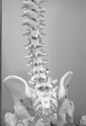
There are 31 pair of spinal nerves. With the exception of C1 all nerves have a ventral
and dorsal nerve root exiting through the intervertebral foramen. This division
somewhat resembles the tongue of a snake as it is bifid. The ventral nerve root
enters the anterior horn while the dorsal nerve root enters the posterior horn.
C1 exits between the cranial occipital bone and 1st cervical vertebra, and
frequently does not have a dorsal root. C2 exits between the axis and atlas (or
between C1 & C2). C8 exits between the 7th cervical vertebra and the 1st
thoracic vertebra. The first thoracic nerve exits between T1 & T2. Therefore in
the cervical region the respective nerves exit above the referenced segment with
the exception of C8, where there is not a corresponding vertebra. In the
thoracic, lumbar and sacral regions the reference segmental nerve supply exits
beneath the referenced vertebral segment.
The upper cervical nerves
associate with cranial nerves as referenced in our cranial nerve section.
Additionally they form the cervical plexus. Plexi are a network of nerves. The
spine has five plexi which frequently overlap. The names of these plexi are: the
cervical, brachial, lumbar, sacral and coccygeal. The thoracic spine for the
most part is not involved in the plexi. All
spinal nerves consist of mixed fibers both sensory & motor. This allows for
neurotransmission from the sensory receptors to the spinal cord or afferent
transmission. The dorsal root is responsible for the conducting of sensory
transmission. Efferent transmission is conducted from the spinal cord to the
muscles for motor response as well as for the conducting of the sympathetic &
parasympathetic pathways. This efferent transmission is predominantly conducted
through the ventral root. The dorsal root merges with the posterior horn and the
ventral root merges with the anterior horn. The spinal roots traverse laterally
and merge slightly distal to the dorsal root ganglion to form a spinal nerve. At
which time they traverse through the intervertebral foramen of their respective
segment and form the rami. The rami can either divide or return back towards the
spine to provide nerve supply to the meninges, vertebra and ligaments or move
into the periphery to supply the skin, muscles and viscera. The
sympathetic chain consisting of 22 ganglia emerge from the ventral root and
involve three cervical segments, eleven thoracic segments, four lumbar
segments, and four
sacral segments. And are responsible for elevated heart rate, increased
electrical activity of the brain, deep and rapid breathing, and dilation of
blood vessels, eyes and galvanic skin response. Or in other words, it is the
system which conducts the “fight or flight” mechanism. The
parasympathetic or craniosacral division is more simplistic anatomically than
the sympathetic division of the autonomic nervous system, due to its preganglionic neurons being located in the brain stem and the sacral region.
Its postganglionic neurons are located in close approximation to the organ to be
supplied. While the parasympathetic division is considered to be a supportive
system for the sympathetic, frequently they have an antagonistic relationship.
This antagonistic relationship must be carefully balanced and regulated. The
parasympathetic division is active during rest. This is when it provides
digestion and the conservation of energy, however, should you eat a large meal
and immediately jog, you have thrown these two divisions into direct
opposition. Hilton’s
Law states: “a nerve trunk which supplies the muscles of any given joint also
supplies the muscles which move the joint and the skin over the insertions of
such muscles.”
Based upon this law and supporting EMG studies, we can assume that
underlying dermatomes are residing myotomes and sclerotomes with resulting
sensory and motor dysfunction. Should there be an organic or biomechanical
encroachment or compression affecting the ventral nerve root you would
anticipate autonomic impairment and subsequent viscerotomes. The most obvious
evidence of a dermatome pattern is the lesions produced by herpes zoster. As
this infection predominantly affects the dorsal root ganglia of the thoracic
segments, dermatomal patterns are outlined by defined pain, hyperesthesia and
pustules. However, it can occur at any level of the spine and following the
active state of the lesions post-herpetic neuralgia may be experienced
periodically for years, and is usually predicated by stress and a compromised
autoimmune system. Radiculopathies also follow these pathways with resulting
dysesthesia.
The spinal nerves have overlapping supply thus serving as a protective mechanism
against injury as it pertains to nerve innervation of a given structure or
organ. Segmental supply as referenced below is derived from consistencies from
Gray’s Anatomy, Correlative Neuroanatomy by Waxman & deGroot, Human Anatomy and
Physiology by Dr. Marieb and various EMG studies. The reader should note that
innervation can occur within a segment or two above or below our specific
reference depending upon the individual.
Below are links to data tables containing information about
spinal nerve segment,
plexus,
innervation of muscle and visceria,
and dermatome pattern for each spinal region.
C1 through C8 - the cervical region
T1 through T12 - the thoracic region
L1 through L5 - the lumbar region
S1 through S5 + coccyx - the sacro-coccygeal region © Copyright American Academy of Manual Medicine. 2001-
. All rights reserved.
Spinal Nerve Supply